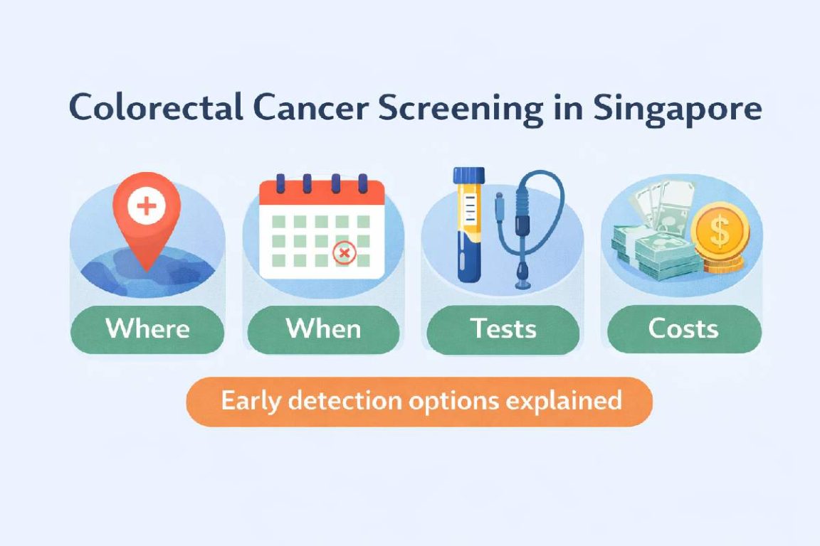
Did you know that colorectal cancer develops over 10-15 years, giving you a substantial window to prevent it entirely? Singapore colorectal cancer screening detects precancerous polyps before they become cancerous or identifies cancer at early, treatable stages. The Ministry of Health recommends screening starting at age 50 for average-risk individuals, with earlier screening for those with family history or genetic conditions.
Three screening methods detect colorectal abnormalities with varying accuracy and invasiveness:
- Colonoscopy visualizes the entire colon directly and removes polyps during the same procedure.
- Faecal immunochemical tests (FIT) detect hidden blood in stool samples collected at home.
- CT colonography uses specialized imaging to create detailed colon images without inserting a scope.
Colonoscopy Screening
Colonoscopy is a widely used method for colorectal cancer detection, allowing direct visualization of the entire colon lining through a flexible scope with a camera. During the procedure, gastroenterologists or a colonoscopy doctor can remove polyps immediately, preventing their progression to cancer. The procedure takes 20-30 minutes under sedation, with most patients experiencing minimal discomfort.
Preparation begins two days before the procedure with dietary restrictions, avoiding high-fiber foods, nuts, and seeds. The day before colonoscopy, patients consume only clear liquids and drink prescribed bowel preparation solutions to clean the colon completely. Common preparations include polyethylene glycol solutions taken in split doses – half the evening before and half the morning of the procedure.
Polyp detection rates during colonoscopy depend on bowel preparation quality and examination technique. Adenomatous polyps, which can become cancerous, appear in various sizes and shapes:
- Sessile (flat)
- Pedunculated (on a stalk)
- Lateral spreading
Advanced adenomas measuring over 10mm or containing villous features require closer surveillance intervals.
After colonoscopy, surveillance intervals depend on findings:
- Clean colonoscopies in average-risk individuals repeat every 10 years
- Finding 1-2 small adenomas requires repeat colonoscopy in 5-10 years
- Three or more adenomas, large adenomas over 10mm, or adenomas with advanced features need surveillance in 3 years
- Patients with numerous polyps may require genetic testing for hereditary syndromes
Faecal Immunochemical Test (FIT)
FIT screening detects microscopic blood in stool samples through antibodies specific to human hemoglobin, offering a non-invasive screening option performed at home. The test requires no dietary restrictions or medication changes before collection, unlike older guaiac-based tests. Patients collect a small stool sample using the provided kit and return it to the laboratory within specified timeframes.
Sample collection involves using the stick provided to scrape stool surface in multiple areas, ensuring representative sampling. The specimen container contains buffer solution that preserves hemoglobin for accurate testing. Laboratories process samples using automated analyzers that measure hemoglobin concentration, with cutoff values typically set at 100 nanograms per milliliter in Singapore.
Positive FIT results require colonoscopy follow-up to identify bleeding sources, which may include:
- Polyps
- Cancer
- Hemorrhoids
- Inflammatory conditions
Annual FIT screening provides cumulative detection benefits when performed consistently.
FIT limitations include inability to detect non-bleeding polyps and potential false positives from benign bleeding sources. Certain medications like aspirin or NSAIDs don’t require discontinuation before FIT, though they may increase false-positive rates. Vitamin C supplements should be avoided three days before testing as they can cause false-negative results.
CT Colonography
CT colonography, also called virtual colonoscopy, uses computed tomography scanning to create detailed 3D images of the colon and rectum. The procedure requires bowel preparation similar to colonoscopy but involves no sedation, allowing patients to resume normal activities immediately afterward. During scanning, a small tube inserted into the rectum gently inflates the colon with carbon dioxide for better visualization.
Image acquisition occurs in two positions – supine and prone – to ensure complete colon visualization and distinguish between stool and polyps. CT scanners complete imaging in under 10 minutes using low-dose radiation protocols. Computer-aided detection software assists radiologists in identifying polyps by highlighting suspicious areas on reconstructed images.
Polyps 6mm or larger detected on CT colonography typically require colonoscopy for removal. Smaller polyps may undergo surveillance with repeat CT colonography in 3-5 years, as their cancer risk remains minimal. Extracolonic findings occur frequently, including benign liver cysts, kidney stones, or aortic aneurysms requiring clinical correlation.
CT colonography proves useful for patients with incomplete colonoscopy due to tortuous anatomy or strictures. Anticoagulation therapy doesn’t require interruption for CT colonography, benefiting patients at high thrombotic risk. However, the procedure cannot remove detected polyps, requiring subsequent colonoscopy for therapeutic intervention.
Important Note
Retained fluid or stool can obscure polyps on CT colonography. Tagging agents mixed with meals before the procedure help distinguish residual material from true polyps through density differences on imaging.
Risk Stratification and Screening Intervals
Average-risk individuals begin Singapore colorectal cancer screening at age 50 with either annual FIT or colonoscopy every 10 years. Those with first-degree relatives diagnosed with colorectal cancer or advanced adenomas before age 60 should start screening at age 40 or 10 years before the youngest diagnosis in the family. Multiple affected relatives or hereditary syndromes require specialized surveillance protocols.
Lynch syndrome carriers face elevated colorectal cancer risk requiring colonoscopy every 1-2 years starting at age 20-25. Familial adenomatous polyposis (FAP) patients develop numerous polyps by adolescence, necessitating annual flexible sigmoidoscopy from age 10-12. Genetic testing identifies at-risk family members who benefit from intensive surveillance.
Inflammatory bowel disease patients with extensive colitis for over 8 years need surveillance colonoscopy with chromoendoscopy every 1-3 years based on risk factors. Previous colorectal cancer or advanced adenoma removal requires enhanced surveillance with colonoscopy at 1 year, then 3 years, then 5 years if no recurrence occurs.
Personal history factors modify screening recommendations beyond family history. Previous pelvic radiation for other cancers increases colorectal cancer risk, warranting earlier screening initiation. Acromegaly patients require colonoscopy at diagnosis and enhanced surveillance based on initial findings.
Preparation and Recovery
Bowel preparation quality directly impacts polyp detection rates during both colonoscopy and CT colonography. Split-dose preparation, consuming half the solution the evening before and half the morning of the procedure, achieves improved cleansing compared to single-dose regimens. Low-volume preparations using sodium sulfate or magnesium citrate offer alternatives for patients unable to tolerate large-volume polyethylene glycol solutions.
Clear liquid diet the day before procedures includes water, clear broth, apple juice, black coffee, and sports drinks without red or purple coloring. Avoiding iron supplements one week before procedures prevents dark stool that obscures visualization. Diabetic patients require medication adjustments, particularly for insulin and SGLT2 inhibitors, coordinated with their endocrinologist.
Post-colonoscopy recovery involves monitoring in the recovery area for 30-60 minutes until sedation effects resolve. Mild bloating and cramping from residual air typically resolve within hours. Dietary progression starts with light meals, avoiding heavy or spicy foods initially. Driving restrictions apply for 24 hours after sedation, requiring arranged transportation home.
Polypectomy sites may bleed up to 14 days post-procedure, particularly with larger polyp removal. Warning signs requiring immediate medical attention include:
- Severe abdominal pain
- Fever
- Heavy rectal bleeding with clots
Anticoagulation resumption timing depends on polyp size and bleeding risk, typically 24-72 hours for small polyps.
Quick Tip
Chilling bowel preparation solutions and drinking through a straw positioned toward the back of the mouth reduces taste perception, improving tolerability.
What Our Colorectal Surgeon Says
Polyp characteristics observed during colonoscopy guide immediate management decisions and future surveillance planning. Paris classification describes polyp morphology – protruding lesions like pedunculated or sessile polyps remove easily with standard polypectomy techniques. Flat or depressed lesions require resection techniques like endoscopic mucosal resection or submucosal dissection.
Optical diagnosis using narrow-band imaging or chromoendoscopy predicts polyp histology during colonoscopy, allowing “resect and discard” strategies for small polyps. NICE classification distinguishes hyperplastic from adenomatous polyps based on color, vessel pattern, and surface characteristics. This reduces pathology costs while maintaining surveillance interval accuracy.
Quality colonoscopy requires adequate withdrawal time, examining the colon systematically while withdrawing the scope. Retroflexion in the rectum and right colon improves adenoma detection behind folds. Position changes during examination help visualize areas hidden by fluid or colonic flexures.
Patient factors affecting colonoscopy quality include body habitus, previous abdominal surgery creating adhesions, and colonic redundancy. Diverticulosis may obscure flat lesions within diverticular segments. These challenges highlight the importance of endoscopists familiar with techniques for complete examination.
Commonly Asked Questions
How painful is colonoscopy with sedation?
Most patients experience minimal discomfort with conscious sedation using midazolam and fentanyl. You may feel pressure or cramping when the scope navigates colonic bends, but severe pain remains rare. Some patients choose to undergo colonoscopy without sedation, experiencing more cramping but avoiding sedation risks and recovery time.
Can I continue aspirin before colonoscopy?
Aspirin continuation during colonoscopy generally remains safe for most polyp removals. Your healthcare professional evaluates bleeding risk based on polyp size and location. High-risk lesions may require aspirin cessation 5-7 days before the procedure. Dual antiplatelet therapy or anticoagulation requires individualized risk assessment balancing thrombotic and bleeding risks.
What happens if polyps are found during screening?
Detected polyps undergo immediate removal during colonoscopy when technically feasible. The removed tissue undergoes histopathological examination determining polyp type – hyperplastic, adenomatous, or serrated. Adenomatous polyps and serrated lesions carry malignant potential, requiring surveillance colonoscopy based on number, size, and histological features.
How accurate is FIT compared to colonoscopy?
FIT detects colorectal cancer with high sensitivity but lower sensitivity for advanced adenomas compared to colonoscopy. Annual FIT screening over multiple years improves cumulative detection rates. Positive FIT results require colonoscopy confirmation, as bleeding may originate from hemorrhoids or upper gastrointestinal sources rather than colorectal neoplasia.
When should screening stop?
Colorectal cancer screening typically continues until age 75 for healthy individuals, with individualized decisions for ages 76-85 based on life expectancy and previous screening results. Patients with limited life expectancy under 10 years due to comorbidities derive minimal benefit from continued screening. Previous negative screening colonoscopies reduce future cancer risk, supporting screening cessation decisions.
Next Steps
Begin screening at age 50 if you’re at average risk, or earlier if you have a family history of colorectal cancer. Choose colonoscopy for direct visualization and immediate polyp removal, annual FIT for non-invasive home testing, or CT colonography if you cannot tolerate traditional colonoscopy. Follow surveillance intervals based on your initial findings to maintain long-term protection.
If you’re experiencing rectal bleeding, changes in bowel habits, or persistent abdominal symptoms, consult a colorectal surgeon for comprehensive evaluation and screening.